Wednesday, December 3, 2008
Buy Dunlop Volleys Nz
HEX400. Picture of the cortex which took in the presence of some pyramidal cells, as shown by the arrow (Larger) with eosinophilic cytoplasm and where you see a small way of axon extension. Looking at other smaller pyramidal cells and the presence of small capillaries in the living tissue or glia with nuclei of oligodendrocytes.
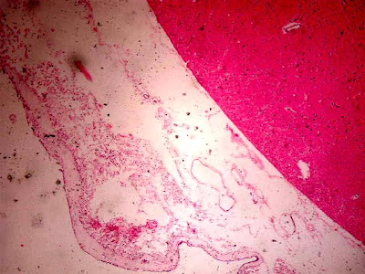
HEX40. Cortex at the peripheral level where we see on the outside deck at its side parietal meningeal (external) or call dura coated monolayer cells. Note the intermediate space with numerous dilated blood vessels and arachnoid membrane and a slight barely noticeable in this expansion or pia mater. Underlying the brain parenchyma.
HEX100. An approach in a field of cortex at the peripheral level where we see on the outside (upper area mage) in the meningeal covering both rock (external) or call dura coated monolayer cells. Note the space between with some dilated blood vessels and arachnoid membrane and a slight barely noticeable in this expansion or pia mater. Suyacente the brain parenchyma where there are aspects of the first cell layers of the cortex.
HEX40. Cortex at Peripheral where we see on the outside (upper area mage) and meningeal covering the brain parenchyma homogeneous and where you can see aspects of their layers seen in this enlargement.
HEX400.
Another detail of the image of the cortex where they took in the presence of some pyramidal cells with eosinophilic cytoplasm and where you see a small way of axon extension in all cases.
presence of small capillaries in the living tissue or glia with nuclei of oligodendrocytes, small and strongly basophilic.
HEX400.
Presence of a dilated capillary in supporting or glial tissue with many nuclei of oligodendrocytes, small and strongly basophilic.
HEX400. Another detail of the image of the cortex where they took in the presence of some pyramidal cells with eosinophilic cytoplasm.
There are other smaller pyramidal cells and the presence of small capillaries and some extravasation of erythrocytes (very common in autopsies) in tissue supporting or glial cells with nuclei of oligodendrocytes.
HEX400.
Another approach detail Image of the cortex where they took in the presence of abundant pyramidal cells in the image together with eosinophilic cytoplasm.
There are other smaller pyramidal cells and the presence of small capillaries and some very focal extravasation of red cells in living tissue or glia with nuclei of oligodendrocytes.
HEX400. Periventricular zone (IV ventricle).
Detail ependymal cells lining with cubic and centrally located nuclei and basophilic appearance. The cytoplasm is finely granular. Within the coating is even observe epndimario numerous small capillaries.
HE X400. An approach to peripheral cortex level den where we see on the outside deck at its side parietal meningeal (outer) on the upper right or call dura coated monolayer cells. Note the space between with some dilated blood vessels and arachnoid membrane and a slight barely noticeable in this expansion or pia mater. Underlying brain parenchyma with some nuclei of glia or oligodendrocytes.
HE X400. An intermediate zone of the cortex which is mainly observed nuclei of oligodendrocytes, glia or substance.
Warranty On Hamilton Beach Hb-p90d23al-dj
Introduction to Nervous System Histology Histology of the cerebral cortex
The presentation of this site is intended primarily for students of the Faculty of Health Sciences in the first cycle and initiate their knowledge of human histology. This page is very simple and basic knowledge of the histology transmitted through simple and original images taken by me. The aim is to interpret images of human histology technique "Standard" color of hematoxylin-eosin (HE). All images are annotated and have a brief pre-theoretical description more understandable to the display of subsequent images. I hope they can serve and bring some knowledge to the virtual community of Histology. All you join I hope you enjoy and make any comments that you may believe appropriate. Thanks for helping.
Greetings to all.
Brazilian Waxing In Portsmouth New Hampshire
Histology Histology of the Cerebellum Spinal Cord
Spinal anterior horn. HEX40.
histological picture anterior horn showing homogeneity of the tissue where there are some cells that correspond to the bodies of previous motor neurons.
Marrow. Previous HEx400.
parenchyma of the anterior horn of the spinal cord showing motor neuron CURP composed of a granular cytoplasm with numerous clusters called Nissl bodies.
Marrow. Previous HEx400.
Another detail of the parenchyma of the anterior horn of the spinal cord showing motoneurons bodies composed of a granular cytoplasm with numerous clusters called Nissl bodies. We can observe the glial support their nuclei of oligodendrocytes.
Details medullary parenchyma in the center where we observe the presence or ependymal cells lining the spinal canal. You can see the consistency of the tissue and the presence of numerous blood vessels, some of them dilated.
enlarged image taken from the back of the bone and the and external surface area extrameníngea. Septation is observed in the lymph node parenchyma and the presence of ganglion cells with vacuolated cytoplasm and basophilic nuclei containing well-defined and fine granulations.
spinal ganglion. HEX400.
image taken from the back of the bone and the external surface area and extrameníngea. The meninges are in the bottom right of the image.
capsular linings are even observe that perfectly delimited.
Any other detail of the parenchyma of the anterior horn of the spinal cord showing the bodies of two motoneurons consist of a granular cytoplasm with numerous conglomerates (Nissl bodies described above).
We can observe the abundant glial support their nuclei of oligodendrocytes.
Spinal HEX400.
ependymal cells.
These cells are also part of the cellular component of glia are cuboid cell cilia in the image can not be appreciated.
These cells contain the nuclei slightly basophilic.
Indianapolis Indiana Drivers License Template
Cerebellum. HEX40. Court which is seen in the center of the image and as highlighted the presence of meningeal cover in this area called the tentorium, but with the same layers as in the rest of the brain parenchyma. Is observed as the arachnoid contains some blood vessels. On the outskirts of Pictured in the parenchyma of the cerebellum.
molecular layer of the cerebellum. HEX400.
This layer is homogeneous in terms of the parenchyma and the presence of matter took in supporting or glial cells with nuclei of oligodendrocytes.
Cerebellum. HEX400. Molecular-granular transition where the top part left is the granular cell layer that define the presence of numerous strongly basophilic nuclear clusters.
2 is even observe Purkinje cells in the intemedia.
Cerebellum X40. Transition zone and granular and molecular layer where you can see the Purkinje cells interspersed between these 2 layers as described above.
Cerebellum. HEX40. Another detail of the Court which is seen in the center of the image and how important the presence of meningeal cover in this area called the tentorium, but with the same layers as in the rest of the brain parenchyma. Is observed as the arachnoid contains some blood vessels, some congestive
In the periphery of the image to see the parenchyma with cerebellar molecular and granular layers.
HEX100 Cerebellum.
Details of the molecular-granular transition, typical of cerebellar parenchyma detail.
At the bottom right, we noticed the tentorium.
Detail obrvmo transition where molecular-granular and three conpuestas purkinje cells by well-defined cytoplasm finely aspect yamplio granular core round and strongly basophilic.
Detail of the granular layer composed of the presence of numerous strongly basophilic nuclear clusters. Ell within dilated capillaries were observed with erythrocytes inside. Glialde fabric is observanzonas support in the center and the image and whose cores correspond to oligodendrocytes.
Subscribe to:
Posts (Atom)