HEX400. Picture of the cortex which took in the presence of some pyramidal cells, as shown by the arrow (Larger) with eosinophilic cytoplasm and where you see a small way of axon extension. Looking at other smaller pyramidal cells and the presence of small capillaries in the living tissue or glia with nuclei of oligodendrocytes.

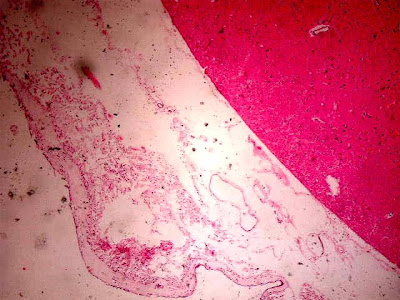
HEX40. Cortex at the peripheral level where we see on the outside deck at its side parietal meningeal (external) or call dura coated monolayer cells. Note the intermediate space with numerous dilated blood vessels and arachnoid membrane and a slight barely noticeable in this expansion or pia mater. Underlying the brain parenchyma.

HEX100. An approach in a field of cortex at the peripheral level where we see on the outside (upper area mage) in the meningeal covering both rock (external) or call dura coated monolayer cells. Note the space between with some dilated blood vessels and arachnoid membrane and a slight barely noticeable in this expansion or pia mater. Suyacente the brain parenchyma where there are aspects of the first cell layers of the cortex.

HEX40. Cortex at Peripheral where we see on the outside (upper area mage) and meningeal covering the brain parenchyma homogeneous and where you can see aspects of their layers seen in this enlargement.

HEX400.
Another detail of the image of the cortex where they took in the presence of some pyramidal cells with eosinophilic cytoplasm and where you see a small way of axon extension in all cases.
presence of small capillaries in the living tissue or glia with nuclei of oligodendrocytes, small and strongly basophilic.

HEX400.
Presence of a dilated capillary in supporting or glial tissue with many nuclei of oligodendrocytes, small and strongly basophilic.

HEX400. Another detail of the image of the cortex where they took in the presence of some pyramidal cells with eosinophilic cytoplasm.
There are other smaller pyramidal cells and the presence of small capillaries and some extravasation of erythrocytes (very common in autopsies) in tissue supporting or glial cells with nuclei of oligodendrocytes.

HEX400.
Another approach detail Image of the cortex where they took in the presence of abundant pyramidal cells in the image together with eosinophilic cytoplasm.
There are other smaller pyramidal cells and the presence of small capillaries and some very focal extravasation of red cells in living tissue or glia with nuclei of oligodendrocytes.

HEX400. Periventricular zone (IV ventricle).
Detail ependymal cells lining with cubic and centrally located nuclei and basophilic appearance. The cytoplasm is finely granular. Within the coating is even observe epndimario numerous small capillaries.

HE X400. An approach to peripheral cortex level den where we see on the outside deck at its side parietal meningeal (outer) on the upper right or call dura coated monolayer cells. Note the space between with some dilated blood vessels and arachnoid membrane and a slight barely noticeable in this expansion or pia mater. Underlying brain parenchyma with some nuclei of glia or oligodendrocytes.

HE X400. An intermediate zone of the cortex which is mainly observed nuclei of oligodendrocytes, glia or substance.
0 comments:
Post a Comment